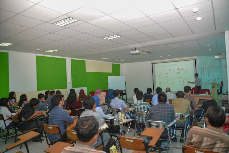
Ruang 203 Gedung Engineering Center FTUI, Indonesia
4th September 2017
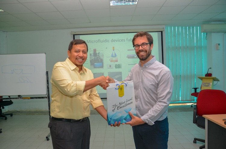
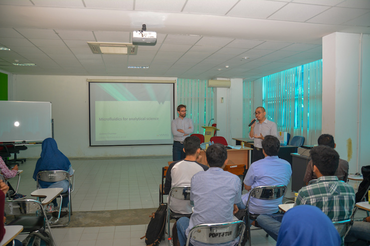
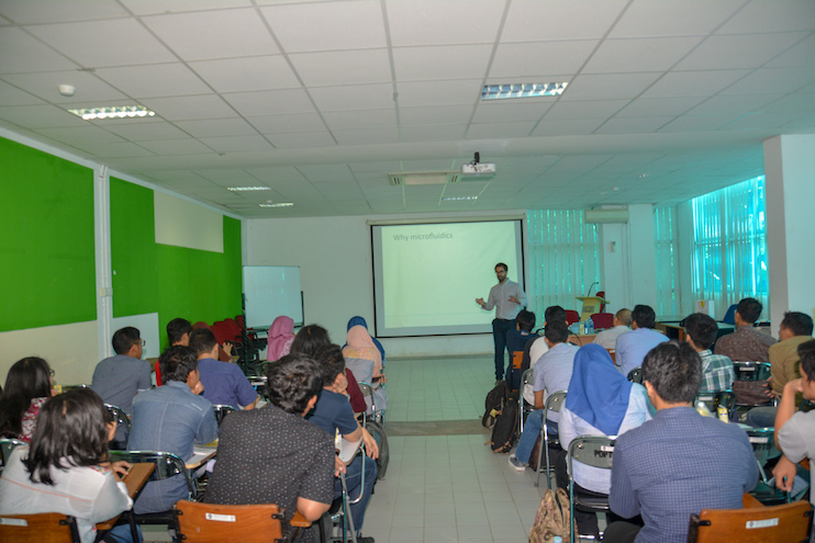
Guest Lecture by Dr. Jerome Charmet from Warwick University
Title: Microfluidics for Analytical Science
Opening and introduction by Dr. Yudan Whulanza
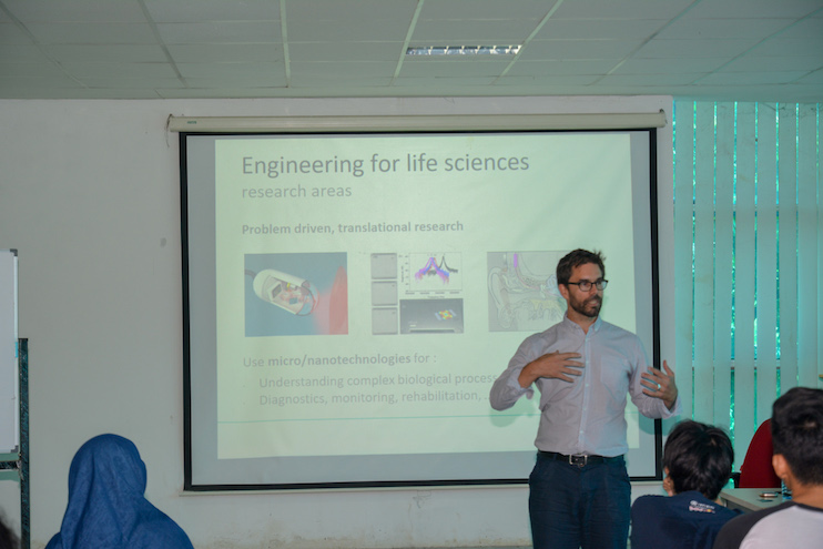
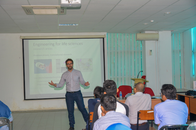
Dr. Jerome Charmet comes here to discuss collaborative work for application of Newton Fund. He is originally from Swiss and had visited Makassar for one year in early 2000s. His current position is associate professor in Warwick University, Coventry, UK with work experience in Intel. His research areas are engineering for life science, micro/nanotechnologies, biosensors, diagnostics, monitoring, and rehabilitation. To perform research, he develops tools including microfabrication, microfluidics, micro/nano-sensors and actuators. His research is in scope of length diagram from microns to nanoscale.
Microfluidics has several features such as flow properties, microenvironment control, and integration. while some governing equations in microfluidics include: reynolds number, peclet number, capillary number, and navier-stokes equation. In microfluidics, we can observe phenomena under laminar flow where diffusion occurs slowly. one way to build microenvironments is through droplet microfluidics. Microfluidic components then can be integrated into lab on a chip.
Dr. Jerome Charmet is currently focusing on using microfluidics to investigate Alzheimer’s disease. Although there is no cure for now, there is an opportunity to manage Alzheimer’s disease since there are anatomically changes 15 years before cognitive decline. Current cause of Alzheimer’s disease is that brain’s amyloids form chain of plaques which then disrupt cerebral blood vessel. The protein structure in brain ranges from monomers, dimers, oligomers, protofibrils, to fibrils.
Beside Alzheimer’s disease there are another protein misfolding and aggregation diseases including Parkinson’s disease, Huntington’s disease, and Diabetes type II. The tools to study protein misfolding are divide into two: single molecule (AFM, TIRF) and bulk measurement (CD, NMR). There are four levels of protein structure but misfolding usually occurs at transition from alpha helix to beta sheet.
Dr. Jerome Charmet developed a technology integrating circular dichroism and microfluidics to perform size separation without losing temporal information and native environment using microfluidics by concentration gradient and diffusion channel. The technique enables separation for bulk information on resolved fractions.
Dr. Jerome Charmet develops a custom soft photolithography machine using UV-LED 365nm, lens, quartz plate, and raspberry. He also develops technique to stabilize liquid structures using parylene through gas deposition to solid on liquid. This can be used to encapsulate functional liquids. One application of this technique is ionic liquid for thermoelectric generator.
Questions from participant:
- From Safira about pdms surface activation using plasma.
- From Sahlan about next step on alzheimer’s research, aggregation and identification of protein, and interaction between inner and outer environment on encapsulated liquid.
- From Zahra about simulation to predict phenomena during experiment.
- From Rizki about nanofluids fabrication.
- From Ardiyansyah about metal as material for microfluidics for heat exchanger.